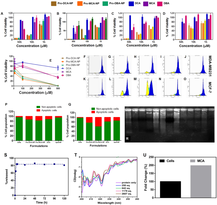
Figure 4. Cell growth inhibition studied by MTT assay.
Assay performed in (A) Panc1; (B) MCF-7; (C) MDA-MB231 and (D) BT549 cells treated with formulations at concentration ranging from 10–500 μM for 48 h and (E) BT549 treated for 72 h. Representative histograms from propidium iodide stained MDA-MB231 and MCF-7 (F-O) for quantifying % apoptotic population (yellow) after treatment with (G,L) Pro-DCA-NP; (H,M) Pro-MCA-NP; (I,N) DCA and (J,O) Lipid-NP formulations at 50 μM for 48 h while (F,K) represent non treated cells. The %apoptotic population compared in (P) MDA-MB231 and (Q) MCF-7 cells. (R) Gel electrophoresis performed on fragmented genomic DNA extracted from MCF-7 (Lane 1–6) and MDA-MB231 (Lane 8–13) cells while lane 7 represent 1 kb ladder. Genomic DNA was extracted from cells treated with (Lane 1, 8) Pro-MCA-NP; (Lane 2, 9) DCA; (Lane 3, 10) untreated cells; (Lane 4, 11) DBA; (Lane 5, 12) MCA and (Lane 6, 13) Pro-DCA-NP formulations at 50 μM in MDA-MB231 and 200 μM in MCF-7 due to different IC50 levels in both the cells for 72 h. (S) Drug release study performed on Pro-MCA-NPs against acetate buffer (pH 4.6); (T) circular dichroism performed for PDK2 protein against added MCA and (U) mitochondrial enrichment of MCA calculated by estimating chloride provide significant input on role of MCA after being released from Pro-MCA-NP.

