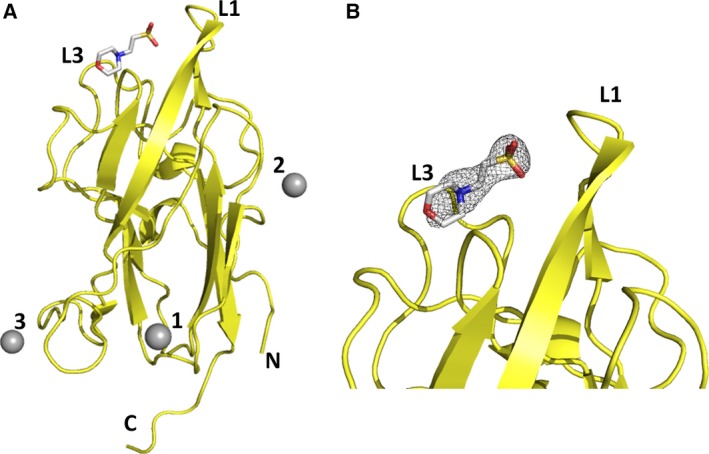
Figure 1.

Zn2+ binding site on NRP2. (A) The X‐ray crystal structure of NRP2 crystallized in the presence of zinc revealed three zinc ions bound to the b1 domain. The structure is shown in yellow ribbon representation, and the three zinc ions are shown as grey spheres. A molecule of MES (2‐ethanesulfonic acid) bound to the canonical ligand binding site on the neuropilin b1 domain is shown using sticks representation. Loops L1 and L3 that shape the binding cleft are indicated. (B) Enlarged view of the ligand binding site, with the MES‐associated 2 F o – F c electron density map shown as a grey wire mesh and contoured at 1 σ.
