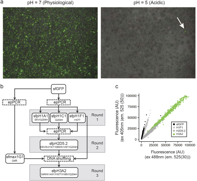
Figure 1. Screening for a pH-stable GFP variant.
(a) Comparison of the fluorescence of nLR-encapsulated cells expressing the GFP library in physiological versus acidic pH. Arrow indicates a potential pH-stable variant. (b) Schematic representation of rounds of mutation and screening to identify a pH-stable GFP variant. (c) Comparison of the ratio of fluorescence intensities at pH 5 of indicated variants at 405 nm and 488 nm with constant emission (525 nm).

