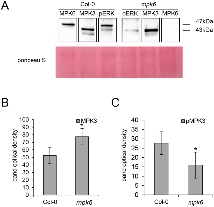
Figure 3. Imunoblotting analysis of MPK3 activity in the mpk6 mutant using phospho-specific pERK antibody.
(A) Representative immunoblots of wild type and mpk6 mutant roots probed with anti-MPK3 (lane MPK3), anti-MPK6 (lane MPK6) and anti-phospho-p44/42 (pERK) antibodies. (B) Quantification of the band optical density corresponding to MPK3 in (A). (C) Quantification of the band optical density corresponding to phosphorylated MPK3 (pMPK3) (band of 43 kDa in lane pERK in A) in wild type and mpk6 mutant roots. Error bars are standard deviations calculated from 3 biological replicates. Stars indicate significant difference at p ≤ 0.05 according to Student t-test.

