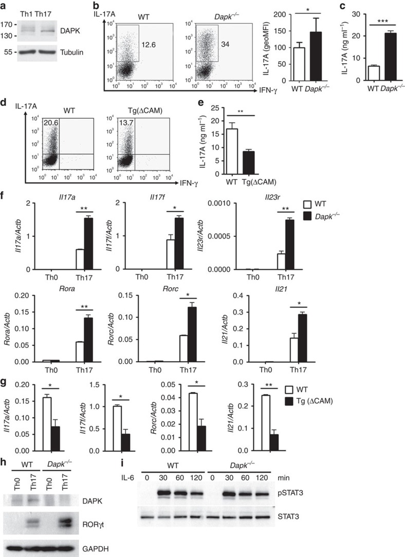
Figure 2. Enhanced Th17 development in Dapk−/− T cells.
(a) Increased DAPK levels in Th17 cells. WT naive CD4 (CD4+CD25−CD44−CD62L+) T cells were subjected to differentiation into Th1 and Th17 cells for 3 days. The levels of DAPK were determined. (b) Increased IL-17 production in Dapk−/− Th17 cells. WT and Dapk−/− Th17 cells differentiated for 5 days were re-stimulated with TPA/A23187 for 4 h in the presence of monensin during the last 5 h, and the expression of IL-17 and IFN-γ were analysed by intracellular staining. The numbers represent the percentages of cells positive (left). The geoMFI is calculated (mean±s.d., n=7) (right). (c) Enhanced IL-17 secretion by Dapk−/− Th17 cells. WT and Dapk−/− Th17 cells were re-stimulated with TPA/A23187 for 24 h, and the levels of IL-17 in the supernatant were measured by ELISA (mean±s.d., n=3). (d,e) The [ΔCAM]DAPK transgene inhibited Th17 differentiation. WT and [ΔCAM]DAPK-transgenic T cells were differentiated into Th17 cells for 3 days. The expression of IL-17 and IFN-γ were determined after restimulation by intracellular staining (d) or by ELISA (mean±s.d., n=3) (e). (f) Elevated expression of Th17-related genes in Dapk−/− T cells. WT and Dapk−/− Th0 and Th17 cells were re-stimulated with TPA/A23187 for 3 h, and RNA was isolated. The expression of Il17a, Il17f, Il21, Il23r, Rorc and Rora were determined by quantitative PCR (mean±s.d., n=2). (g) Attenuated expression of Th17-associated genes in [ΔCAM]DAPK-transgenic T cells. The expression of Il17a, Il17f, Rorc and Il21 in WT and [ΔCAM]DAPK-transgenic Th17 cells were determined as in f. Mean±s.d., n=3. (h) Increased expression of RORγt protein in Dapk−/− Th17 cells. A fraction of the TPA/A23187-re-stimulated cells from b were analysed for RORγt expression by western blot. (i) Normal STAT3 activation in Dapk−/− T cells. Freshly isolated WT and Dapk−/− T cells were stimulated with IL-6 for the indicated times. The levels of phospho-STAT3 and total STAT3 were examined by western blotting. Data (a,c–i) are representative of three independent experiments. *P<0.05, **P<0.01, ***P<0.001 for unpaired t-test.

