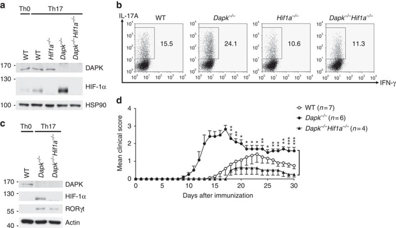
Figure 7. HIF-1α-knockout prevents excess Th17 differentiation in Dapk−/− T cells.
Naive CD4+ T cells from WT, Dapk−/−, Hif1a−/− and Dapk−/−Hif1a−/− mice were allowed to differentiate into Th0 and Th17 cells. (a) The levels of DAPK and HIF-1α were determined in Th17 cells and Th0 cells after 3 days of differentiation. (b,c) HIF-1α deficiency reduces the excess Th17 differentiation in Dapk−/− T cells. TPA/A23187-stimulated production of IL-17A and IFN-γ was examined in Th17 cells after 5 days of differentiation (b), and the contents of HIF-1α and RORγt were determined (c). Data are representative of three (b) or two (a,c) independent experiments. (d) HIF-1α deficiency prevents exacerbated EAE generation in Dapk−/− mice. EAE was induced in WT, Dapk−/−, Hif1a−/− and Dapk−/−Hif1a−/− mice as in Fig. 1a. Mice were monitored for clinical signs of paralysis. Values are mean±s.e.m., *P<0.05, **P<0.01, ***P<0.001 for unpaired t-test.

