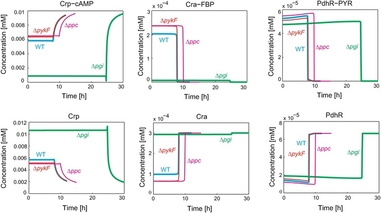
Fig. 3.

TF and TF-metabolite concentration of WT and genetic mutant strains. The blue, red, green and magenta lines indicate WT, ∆pykF, ∆pgi and ∆ppc, respectively

TF and TF-metabolite concentration of WT and genetic mutant strains. The blue, red, green and magenta lines indicate WT, ∆pykF, ∆pgi and ∆ppc, respectively