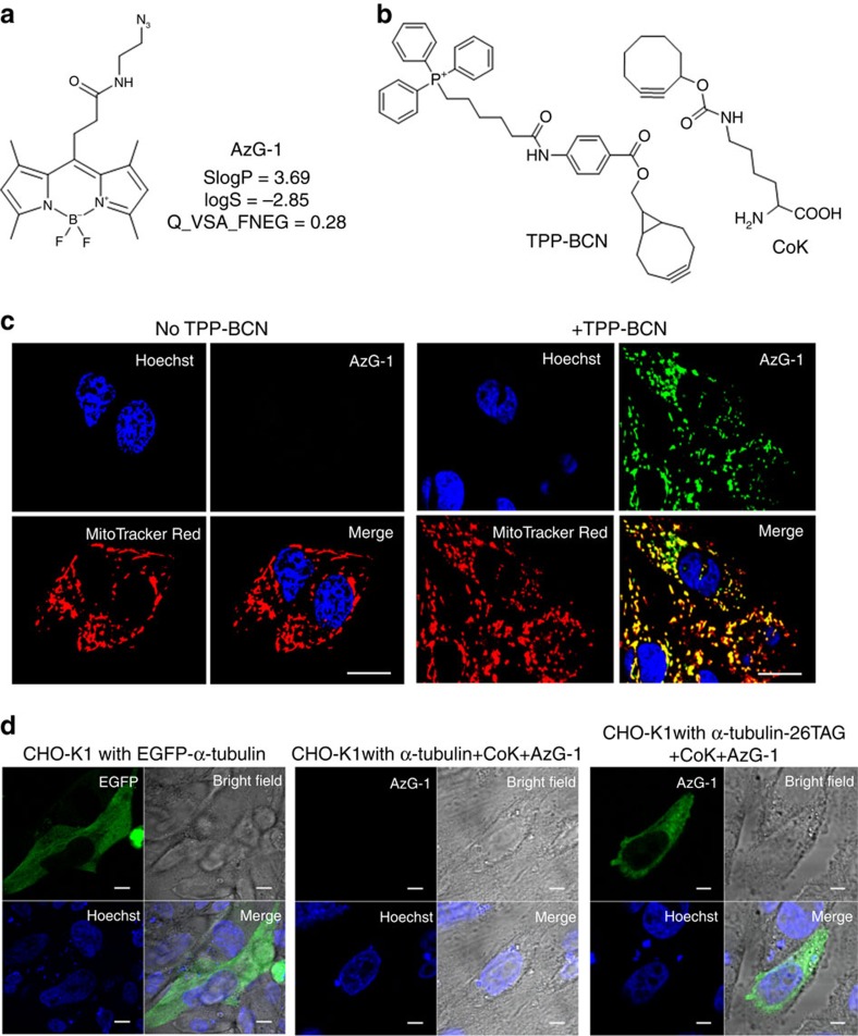
Figure 4. Live cell imaging with AzG-1.
(a) Chemical structures of azide-bearing probe AzG-1 and its descriptor values. (b) Structure of triphenylphosphonium analogues TPP-BCN and cyclooctyne-containing unnatural amino acid, CoK. (c) Fluorescence imaging of mitochondria in U-2 OS cells labelled with AzG-1. Cells were incubated without (left side) and with (right side) TPP-BCN and then labelled with 10 μM AzG-1 for 2 h followed by counterstaining with mitochondria marker. Scale bar, 10 μm. (d) Fluorescence imaging of α-tubulin in live CHO-K1 cells. CHO-K1 cells were co-transfected with plasmids pCoKRS-tRNA and pTub-26TAG to incorporate CoK into α-tubulin at position 26. After incorporation of CoK, cells were labelled with 10 μM AzG-1 for 2 h at 37 °C. CHO-K1 cells were also transfected with plasmid pTubwt to express wild type α-tubulin and treated with CoK and AzG-1 in the same way. CHO-K1 cells transfected with plasmid pEGFP-Tubwt expressing EGFP-fused wild type α-tubulin was used as a control. Live cell images show that AzG-1 labelled mitochondria (c) and is conjugated specifically with CoK-bearing α-tubulin (d). Scale bar, 10 μm.

