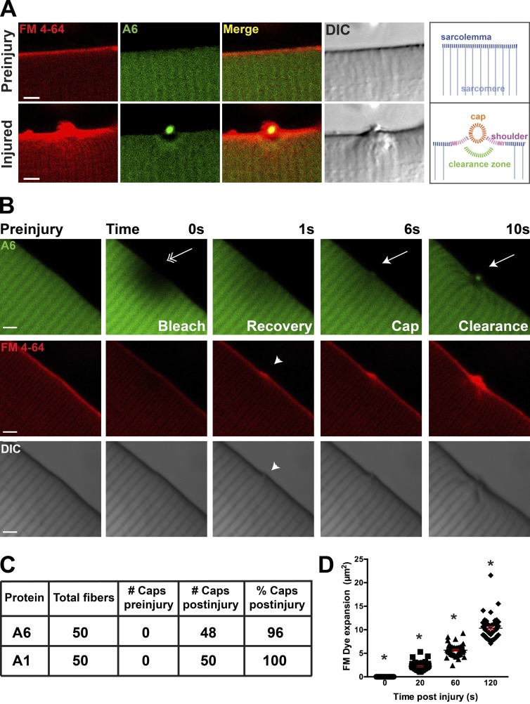
Figure 1.
Annexin A6 forms a repair cap at the site of myofiber sarcolemmal disruption. (A) High-resolution imaging of a single myofiber before laser ablation and then 80 s after sarcolemmal disruption. FM4-64 (red) increased fluorescence after binding lipids, particularly negatively charged lipids, like phosphatidylserine (Zweifach, 2000; Yeung et al., 2009). Upon membrane disruption, FM4-64 accumulated at the site of membrane disruption (bottom, left). Annexin A6 (A6)–GFP (green) was rapidly recruited to the site of FM4-64 aggregation, forming a repair cap after damage (yellow, merge). Within the myofiber, below the annexin A6 cap, was a region devoid of annexin A6 termed the clearance zone. A repair cap and shoulder visualized by differential interference contrast imaging. A schematic is shown indicated with labeling. (B) Laser-injury produced local photobleach that resolved by 1 s postinjury (double arrow). After fluorescence recovery, a repair cap and clearance zone formed (arrow). FM4-64 fluorescence accumulated at the lesion (arrowhead). Bars, 4 µm. (C) Myofibers expressing annexins A6 and A1 produced repair caps in 96% and 100% of damaged fibers, respectively. (D) The technique of laser damage is highly reproducible, as indicated by similar FM4-64 expansion zones after injury at each time point tested (*, P < 0.05; ∼10 myofibers per animal; n = 6 mice per condition). Error bars represent SEM.

