Figure 3.
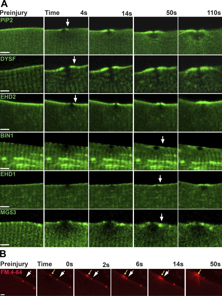
T-Tubule–associated proteins are found at the repair shoulder. (A) PIP2, marked with PLCΔPH, accumulated at the shoulder region and was visualized as early as 4 s after sarcolemmal disruption. Dysferlin (DYSF) and EHD2 were also recruited to the shoulder region within 5–15 s after injury. BIN1, EHD1, and MG53 were recruited between 14 and 50 s after membrane disruption. (B) Aggregates of FM4-64 within the plasma membrane were observed to move laterally along the surface of injured myofibers toward the site of membrane disruption. Injury was conducted in the presence of FM4-64. Fibers containing small, static patches of FM4-64 were selected (white arrow) to provide a point of reference. The static patch of FM4-64 on the sarcolemma moved laterally toward the site of laser damage (yellow arrow) through the plasma membrane. Bars, 4 µm.

