Figure 6.
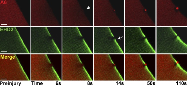
Distinct cap and shoulder repair proteins. Myofibers were coelectroporated with plasmids expressing annexin A6 tagged with mCherry (A6) and EHD2 tagged with GFP (EHD2) to visualize cap and shoulder protein simultaneously. By 8 s postinjury, the annexin A6–rich cap was visualized (arrowhead). The adjacent EHD2-containing shoulder protein was seen adjacent to, but not overlapping with, the A6-rich cap (arrow). The clear zone was devoid of both A6 and EHD2 proteins. Both proteins continued to accumulate through 110 s of imaging. This pattern of localization occurred in 12/12 myofibers collected from four animals. Bars, 4 µm.

