Figure 4.
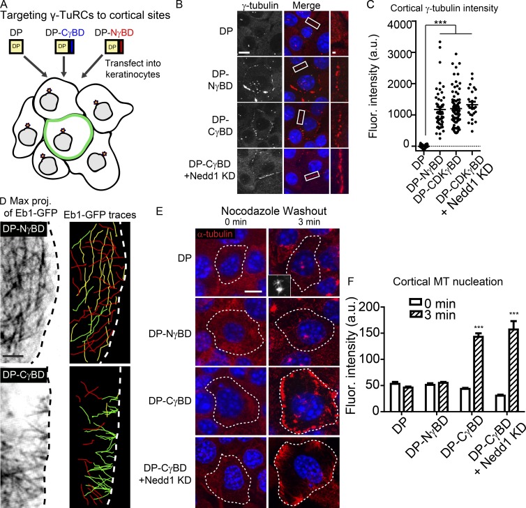
CDK5RAP2, but not Nedd1, is sufficient to stimulate γ-TuRC–mediated MT nucleation in vivo. (A) Diagram of the experimental method. (B) γ-Tubulin localization in cells expressing DP alone, DP-NγBD, DP-CγBD, and DP-CγBD + Nedd1KD. Insets show zoomed in cortical regions. Bars: (main) 10 µm; (insets) 1 µm. (C) Quantification of cortical γ-tubulin levels in cells expressing DP, DP-NγBD, DP-CγBD (all n ≥ 50 cells from three independent experiments), and DP-CγBD + Nedd1KD (n = 25 cells from three independent experiments). (D) Compressions of movies (60 s) of GFP-Eb1 showing MT paths in cells expressing DP-NγBD or DP-CγBD (n ≥ 16 cells from three independent experiments). Images on the right are color coded by vectors of growth. Those growing toward the plasma membrane are red, those growing parallel to it are yellow, and those that are growing into the cytoplasm are green. (E) Representative images of transfected cells before and after nocodazole washout showing sites of MT nucleation. Inset shows centrosomal nucleation in control cells. Dotted line indicates the outline of the transfected cell. Bar, 10 µm. (F) Quantification of cortical α-tubulin intensity in control or DP-NγBD, DP-CγBD, or DP-CγBD + Nedd1KD cells 3 min after nocodazole washout (n = 23–92 cells from at least three independent experiments). ***, P < 0.001. Data are presented as mean ± SEM.

