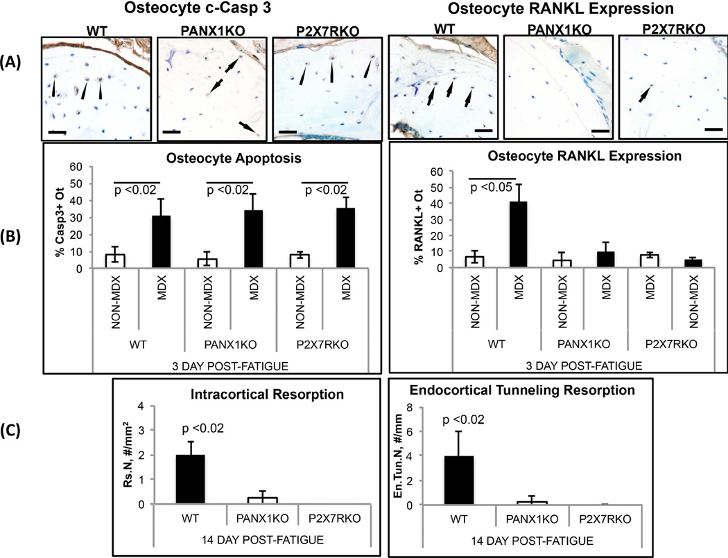
Figure 3.
Osteocyte apoptosis, osteocyte RANKL expression and resorption in fatigued ulnae of WT, Panx1KO and P2X7RKO mice. (A) Left side photomicrographs show IHC staining for osteocyte apoptosis within MDX regions of WT, Panx1KO and P2X7RKO mice at 3 days post-fatigue (bar = 25 µm) (c-Caspase 3 staining); arrows illustrate examples of positively-stained cells (brown colored). Right side photomicrographs show IHC staining for osteocyte RANKL expression in MDX regions at 3 days post-fatigue; RANKL staining (arrows) is evident in WT ulnae, but effectively absent from fatigued ulnae from Panx1KO and P2X7RKO mice. (B) Histomorphometry data for osteocyte apoptosis (%Casp3+ Ot.) and osteocyte RANKL expression (%RANKL+ Ot.) in fatigued ulnae at 3 days after loading: Osteocyte apoptosis at microdamage sites occurred similarly in Panx1KO, P2X7RKO and WT ulnae, consistent with IHC images shown in (A), with apoptosis increased almost 5-fold vs. NON-MDX bone areas with same bone (p<0.02). In contrast, osteocyte RANKL expression was absent from fatigued Panx1KO and P2X7RKO bones. (C) Histomorphometry data for resorption space number (Rs.N) and endocortical tunneling foci number (En.Tun.N) in WT, Panx1KO and P2X7RKO ulnae at 14 days after fatigue (p<0.02 vs WT), showing complete absence of new resorption activity in both KO strains.

