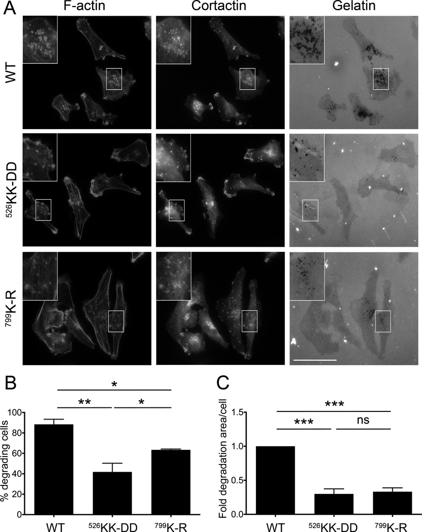
Figure 6. Gβγ binding to p110β is required for gelatin degradation.
A) Representative micrographs of MDA-MB-231 cells expressing wild type or mutant p110β, plated on Oregon Green 488-conjugated gelatin. Cells were stained with rhodamine phalloidin and cortactin to identify invadopodia. B) The percentage of cells with degradative invadopodia. C) The average area of gelatin degradation per cell. Values were normalized to those obtained from cells expressing wild type p110β. Data represent the mean ± SEM from three independent experiments, with a total of 146, 138, and 120 cells for wild type p110β, p110βKK-DD, or p110βK-R, respectively. **: p<0.01; ***: p<0.001.

