. 2016 Apr 30;49(4):201–207. doi: 10.5483/BMBRep.2016.49.4.042

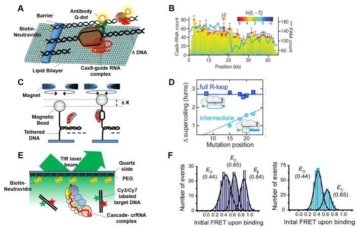
Fig. 2. Various single-molecule approaches for dynamic observation of the CRISPR-Cas system. (A) Tethered DNA curtain-based observation of the movement of a single Cas9 protein on target DNA. Cas9 movement was followed using 3X-FLAG tag-mediated Q-dot labeling. The crRNA-tracrRNA was designed for target sequence (λ2) binding on λ-DNA. (B) Distribution of Cas9–RNA binding events (n = 2,330) and PAM sites. Color-coding indicates the binding dwell time (ti) relative to the mean dwell time (t). Figure taken from (Samuel H. Sternberg et al. Nature, 2014). (C) Magnetic tweezers-based twisting assay. R-loop formation induced by the Cascade complex on negatively supercoiled DNA causes local DNA untwisting. Compensatory overtwisting of the DNA changes the supercoiling, resulting in a change in DNA length (Δx). Each mutant DNA template contains mutations at various locus from the PAM in wild-type DNA template (40). (D) Mean supercoiling changes associated with full (blue) and intermediate R-loop formation (light blue). Mutation position indicates the distance between PAM and mismatch position. Figure taken from (Marius Rutkauskas et al. Cell Reports, 2015). (E) Schematic of smFRET experiment for monitoring Cascade binding to labeled DNA substrates. Dual color (Cy3 and Cy7, shown in green and red star) labeled bona fide target construct contains a 15 bp flank, a PAM, and a protospacer (43). (F) FRET histogram from binding traces of Cascade to bona fide (Left) and PAM-mutated templates (Right) respectively. Figure taken from (Timothy R. Blosser et al. 2015, Mol Cell).

Fig. 2.