. 2016 Apr 30;49(4):214–219. doi: 10.5483/BMBRep.2016.49.4.220

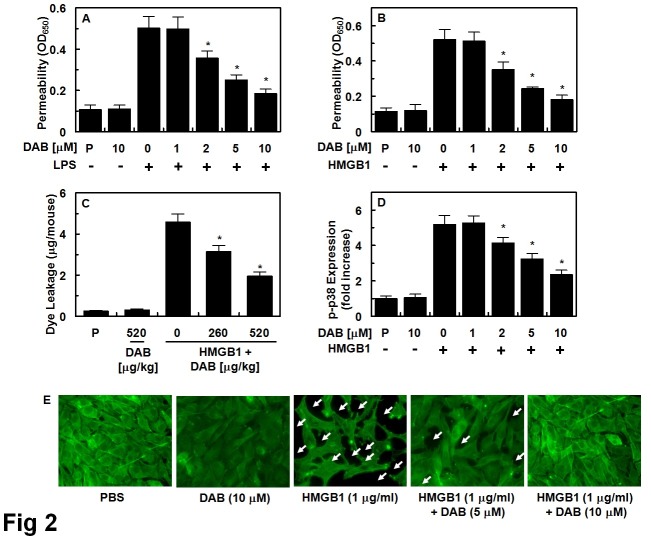
Fig. 2. Effects of DAB on HMGB1-mediated permeability in vitro and in vivo. Effects of treatment with different concentrations of DAB for 6 h on barrier disruption caused by LPS (A, 100 ng/ml, 4 h) or HMGB1 (B, 1 μg/ml, 16 h) were monitored by measuring the flux of Evans blue-bound albumin across HUVECs. (C) The effects of DAB on HMGB1-induced (2 μg/mouse, i.v.) vascular permeability in mice were examined by measuring the amount of Evans blue in peritoneal washings (expressed μg/mouse, n = 5). (D) HUVECs were activated with HMGB1 (1 μg/ml, 16 h), followed by treated with different concentrations of DAB for 6 h. The effects of DAB on HMGB1-mediated expression of phospho-p38 were determined by ELISA. (E) Staining for F-actin. HUVEC monolayers grown on glass coverslips were stimulated with or without HMGB1 for 1 h, and then treated with DAB (5 or 10 μM) for 6 h, and stained for F-actin. Arrows indicate intercellular gaps. The representative images were from three separate experiments in different days with similar results. Results are expressed as the mean ± SEM of three separate experiments in different days. *P < 0.05 versus LPS (A) or HMGB1 (B-D).

Fig. 2.