. 2016 Apr 30;49(4):214–219. doi: 10.5483/BMBRep.2016.49.4.220

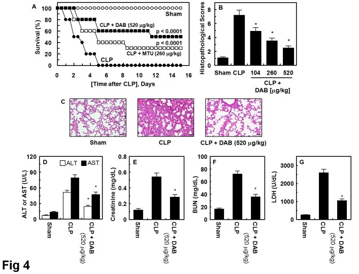
Fig. 4. Effects of DAB on lethality or pulmonary injury after CLP. (A) Male C57BL/6 mice (n = 20) were administered DAB at 260 μg/kg (i.v. □) or at 520 μg/kg (i.v. ■) at 12 h and 50 h after CLP. Animal survival was monitored every 12 hours after CLP for 15 days. Control CLP mice (●) and sham-operated mice (○) were administered sterile saline (n = 20). Kaplan-Meier survival analysis was used to determine the overall survival rates versus CLP treated mice. (B) Male C57BL/6 mice underwent CLP and were administered DAB intravenously at 12 h and 50 h after CLP (n = 5). Mice were euthanized 96 h after CLP. Histopathological scores for the lung tissue were recorded as described in methods. (C) Photomicrographs of lung tissues (H&E staining, ×200). Sham group (grade 1); CLP group (grade 3); Right, CLP + DAB group, (grade 2). Illustrations indicate representative images from three independent experiments in different days with similar results. All results indicate the mean ± SEM of three separate experiments in different days with similar results. (D-G) The same as (B, C) except that mice were bled and euthanized. AST, ALT, BUN, and creatinine level in plasma was measured. All results indicate the mean ± SEM of three separate experiments. *indicates P < 0.05 vs. CLP.

Fig. 4.