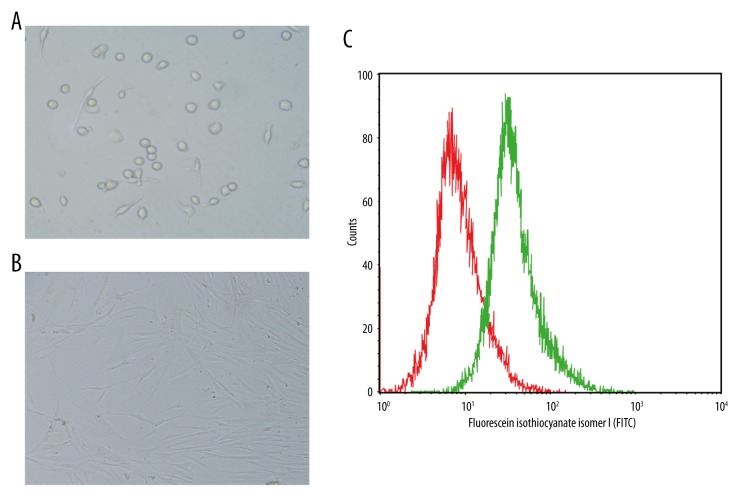
Figure 1.
Isolation and culture of Sca-1+ CSCs and identification of Sca-1+ CSCs by flow cytometry (A – After 1 day of culture, the majority of cells were found to adhere to the wall, with round shapes; B – After 3 days of culture, the majority of cells were spindle-shaped and grew larger; C – identification of Sca-1+ CSCs by flow cytometry).

