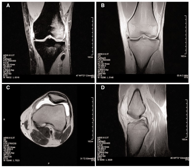
Figure 1.
A 21-year-old male patient with knee trauma. A. Coronal gradient-echo (GE) fat suppressed image shows bone bruise at lateral femoral condyle and lateral tibial plateau as well as turn of MCL. B. Coronal GE in phase T2-weighted image shows normal MM. C. Axial spin-echo (SE) T2 weighted image shows joint effusion. D. Sagittal GE in phase T2-weighted image shows complete tearing of ACL.

