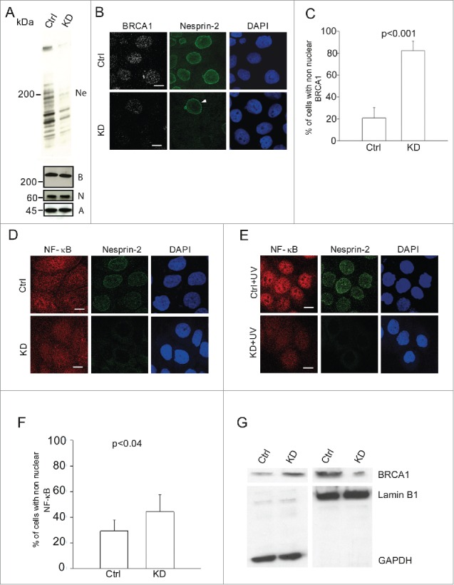
Figure 1.

Reduced nuclear localization of BRCA1 and NF-κB in Nesprin-2 knockdown (KD) HaCaT cells and its effect on UV induced DNA damage response. (A) Western blots showing Nesprin-2 (Ne), BRCA1 (B) and NF-κB (N) levels in whole cell lysates obtained from control (Ctrl) and Nesprin-2 KD cells. Nesprin-2 was detected with pAbK1. β-actin (A) detected with monoclonal antibodies was used for loading control. (B) Immunofluorescence study in control (Ctrl) and Nesprin-2 knockdown (KD) cells using BRCA1 (shown in gray scale) and pAbK1 anti-Nesprin-2 antibodies (shown in green). Arrowhead points at a control cell surrounded by Nesprin-2 KD cells. Scale bar, 10 µM. (C) Graphical representation of the percentage of cells with predominantly non-nuclear BRCA1 (mean ± s.d, p< 0.001). 200 cells were analyzed. (D) Distribution of NF-κB in control and knockdown cells in the absence of UV treatment. Staining was with polyclonal antibodies specific for NF-κB and mAb K20–478 against Nesprin-2. (E) Distribution of NF-κB in control and Nesprin-2 KD cells after UV treatment. Scale bar, 10 µm. (F) Graphical representation of the percentage of cells with predominantly non-nuclear NF-κB post UV treatment (mean ± s.d, p value, 0.0396). 200 cells were analyzed. (G) Subcellular fractionation of control and Nesprin-2 KD cells to reveal the distribution of BRCA1. The resulting blot was probed for BRCA1. Shown are the respective cytosolic (GAPDH positive) and nuclear fractions (Lamin B1 positive).
