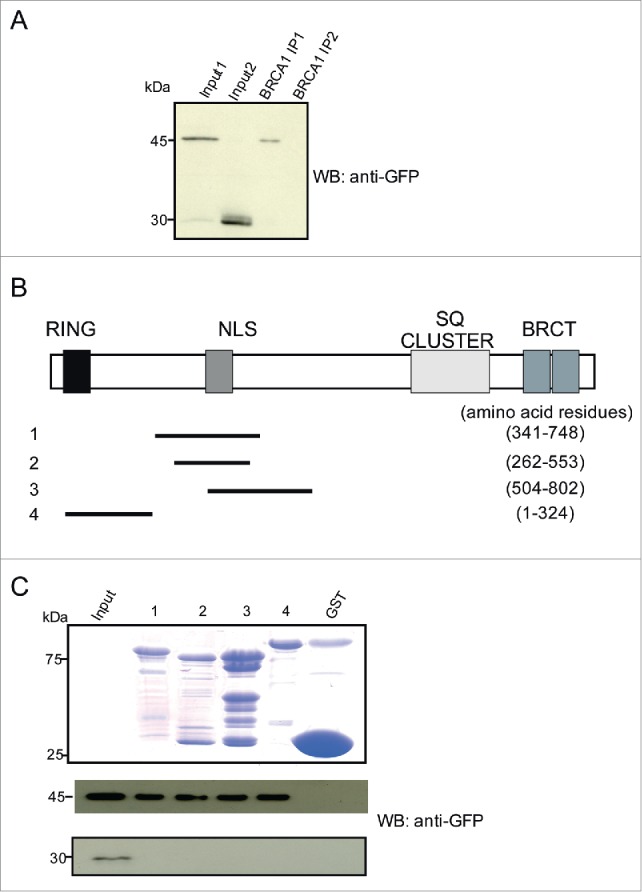
Figure 4.

Calmodulin interacts with BRCA1 and the interaction is mediated via its NLS containing region. (A) Detection of YFP-Calmodulin in the BRCA1 immunoprecipitate. COS7 cells expressing YFP-Calmodulin (~45 kDa) were used. YFP-Calmodulin was detected using GFP-specific mAb K3–184–2. For the pulldown BRCA1 specific antibodies were used. YFP alone was not precipitated. Input 1 and 2 show the respective cell lysates containing YFP-Calmodulin and YFP used for pull down (IP1 and 2, respectively. (B) Position of peptides employed along the 1863 aa BRCA1 protein. The location of the domains of BRCA1 is indicated. (C) Detection of Calmodulin in pull downs using GST fusions of the indicated BRCA1 polypeptides. A Coomassie Blue stained gel shows the GST-fusion proteins. COS7 cells expressing YFP-Calmodulin were used. YFP-Calmodulin was detected by mAb K3–184–2. The lower panel shows the control pull down performed with lysates of cells expressing YFP only.
