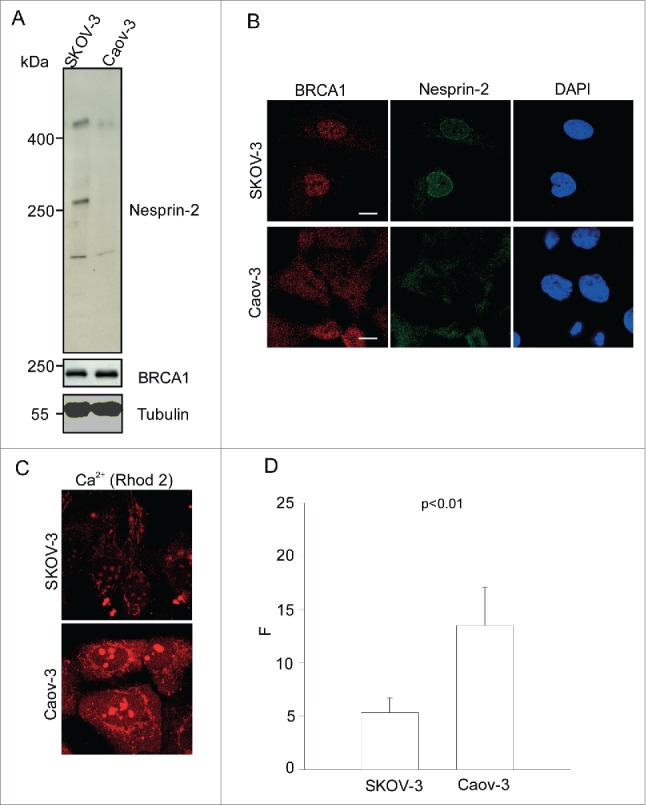
Figure 7.

Nesprin-2 expression, BRCA1 localization and steady-state Ca2+ in ovarian adenocarcinoma cell lines SKOV-3 and Caov-3. (A) Western blot analysis using lysates obtained from SKOV-3 and Caov-3 cells and probed with pAbK1 for Nesprin-2 and BRCA1 antibodies. The proteins were separated in a 3–10%- gradient gel. YL1/2 antibodies were used to detect β-tubulin. (B) Immunofluorescence analysis performed on SKOV-3 and Caov-3 cells using BRCA1 antibodies, Nesprin-2 pAbK1 antibodies and DAPI. (C) Representative images of SKOV-3 and Caov-3 cells loaded with Rhod-2 to detect steady state cytoplasmic Ca2+. (D) Quantitative comparison of fluorescence measurements in the 2 cell lines (F: corrected Rhod-2 fluorescence intensity). The values represent mean and SD of 3 independent experiments (p value, 0.0076). 100 cells were analyzed.
