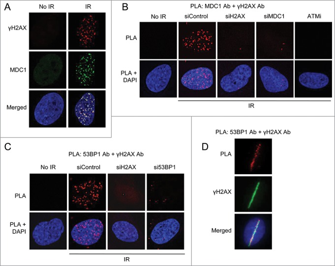
Figure 1.

PLA visualizes complex formation and localization of repair factors at DNA breaks. (A) Immunofluorescent staining of γH2AX, a marker for DNA double-strand breaks, and MDC1 in U2OS cells not treated or whole-cell irradiated (6 Gy, 1 hour recovery). Nuclei were stained with DAPI (in blue) in all immunofluorescence and PLA experiments. (B) PLA detection of MDC1-γH2AX interactions visible as distinct fluorescent dots in U2OS cells. Approximately 90% of non-irradiated cells showed no signals and the remainder 2 dots/cell. In irradiated (6 Gy) cells, 100% of cells displayed >10 dots/cell. U2OS cells were transfected with the indicated siRNAs for 48 hours or treated with ATMi for 16 hours, irradiated and 15 minutes later subjected to PLA using MDC1 and γH2AX antibodies. (C) PLA detection of 53BP1-γH2AX interactions. U2OS cells were transfected with the indicated siRNAs for 48 hours, irradiated (6 Gy) and 15 minutes later subjected to PLA using 53BP1 and γH2AX antibodies. (D) U2OS cells were micro-irradiated and fixed after 5 minutes. PLA was performed to detect 53BP1-γH2AX interactions and cells were counterstained for γH2AX to visualize DNA double-strand breaks.
