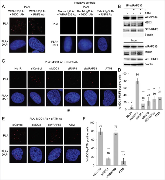
Figure 4.

WRAP53β facilitates MDC1-RNF8 interaction. (A) PLA signals of WRAP53β-MDC1 and WRAP53β-RNF8 interactions in irradiated (6 Gy, 15 minutes recovery) U2OS cells. Negative controls for PLA, showing the detection of MDC1, RNF8 or WRAP53β combined with the indicated normal IgG antibody in irradiated (6 Gy, 15 minutes recovery) U2OS cells. The images show representative numbers of interactions. (B) U2OS cells were either left untreated, irradiated with 6 Gy or treated with ATMi for 16 hours prior to irradiation with 6 Gy. Fifteen minutes later, IP of WRAP53β was performed followed by immunoblotting of WRAP53β, MDC1, GFP-RNF8 and β-actin. (C) PLA signals of MDC1-RNF8 interactions in U2OS cells treated with siControl, siMDC1, siRNF8 or siWRAP53#2 for 48 hours or ATMi for 24 hours, irradiated with 6 Gy and fixed after 15 minutes. (D) Quantification of the results in (C). PLA signals were quantified in 100 cells for each experiment and nuclei containing ≥4 signals were counted as positive cells. The majority of the positive cells showed the same amount of PLA signals per cell as the corresponding positive control (>10 dots/cell), whereas the negative cells mostly had less than 2 dots/cell. (E) PLA signals of MDC1-pATM interactions in U2OS cells treated with siControl, siMDC1, siWRAP53#2 for 48 hours or ATMi for 24 hours, irradiated with 6 Gy and fixed after 15 minutes. (F) Quantification of the results in (E). PLA signals were quantified in 100 cells for each experiment and nuclei containing ≥4 signals were counted as positive cells. Error bars, s.e.m.; n=3, *** p<0.001, Student's t-test.
