
Figure 4.
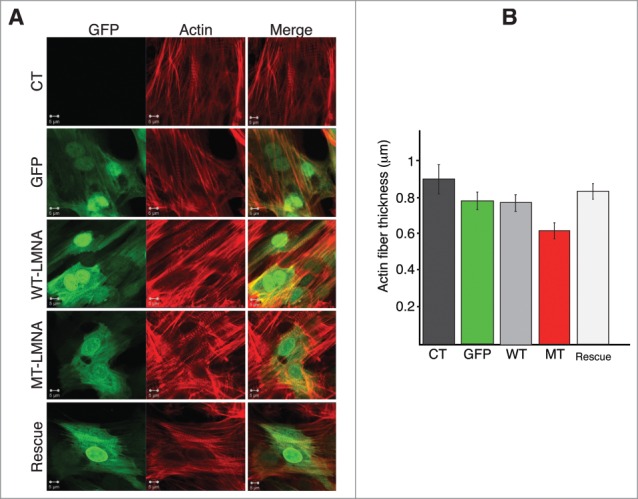
D192G LMNA actin network in NRVMs. (A) Indirect immuofluorescence representative images of NVRMs. Cardiomyocytes expressing mutant LaminA protein display severe decrease of both density and thickness of cytoskeletal actin microfilaments compared to not infected (CT) and NRVMs infected with either WT-LMNA or GFP only. The re-expression of WT Lamin A protein in cardiomyocytes previously infected with MT-LMNA, lead to a restored actin network comparable to controls (Rescue). (B) Quantitative measurement of the actin filaments thickness showed a significant reduction of this cytoskeleton component in MT NRVM compared to both CT and WT cells (p < 0.0001), rescued by the second infection with WT LMNA (p = 0.096 compared to CT).