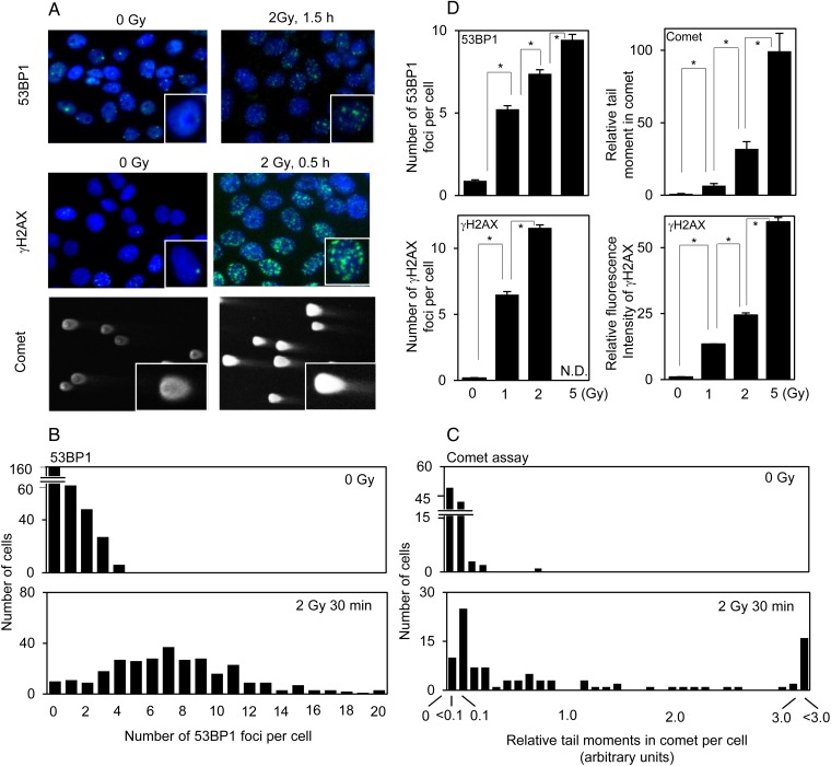
Fig. 1.
DSBs determined by 53BP1/γH2AX staining and the neutral comet assay in control PCCL3 cells and cells irradiated with 2 Gy IR: (A) representative photographs of 53BP1/γH2AX staining and the comet assay; (B and C), the distribution of the number of 53BP1 foci and tail moments in the comet assay, respectively, in control and irradiated cells. (D) effect of IR dose (0 to 5 Gy) on 53BP1/γH2AX staining and the comet assay. *P < 0.01.

