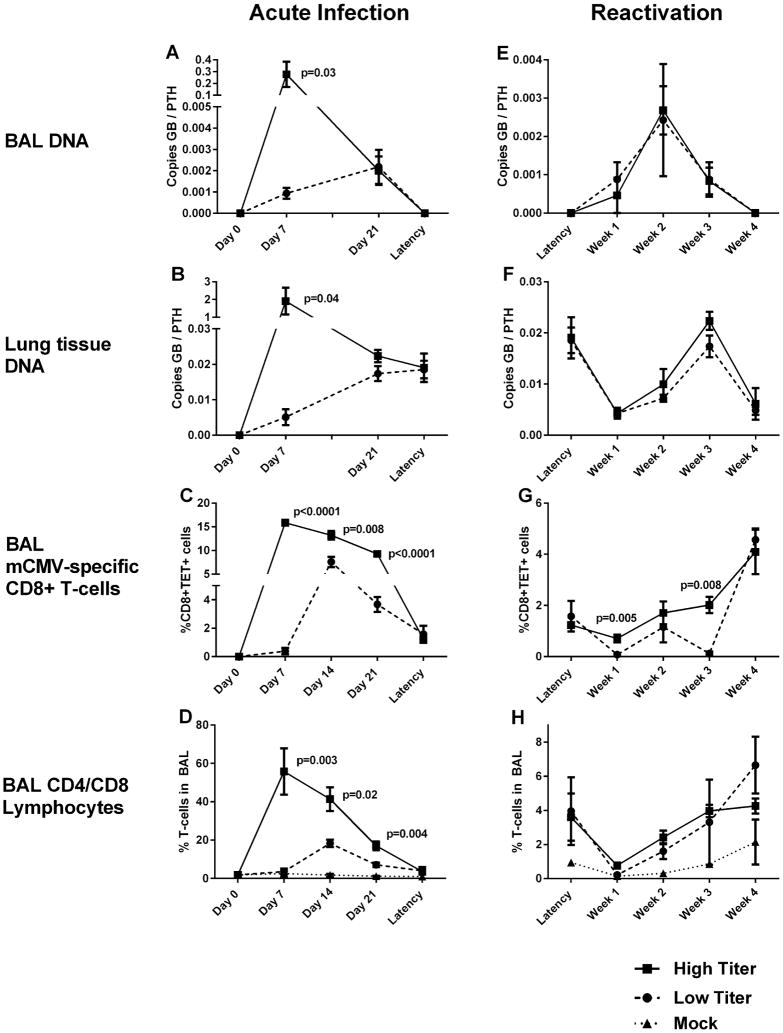
Fig. 1.
Bronchoalveolar lavage (BAL) fluid and lung tissues were collected from euthanized mice infected with murine cytomegalovirus (mCMV) at low titer (1 × 10² plaque forming units (PFU)) and high titer (1 × 10⁶ PFU) initial infections. Time points studied include: 7, 14, and 21 days following acute infection, during latency (4 months after infection), and 1, 2, 3, and 4 weeks following bacterial sepsis from cecal ligation and puncture. Day 0 time-points are prior to infection and mock mice (without infection) are included where applicable. BAL specimens were analyzed by polymerase chain reaction for mCMV-glycoprotein B (GB) relative to control gene parathyroid-related protein (PTHrP) during acute infection (A) and sepsis-induced viral reactivation (E). Similar PCR analyses of lung tissues obtained after BAL collection (B and F). mCMV-specific CD8+ T cells were evaluated via flow cytometry from BAL fluid using tetramers specific for viral peptides m123 or m164 (TET+) during acute infection (C) and sepsis-induced reactivation (G). Overall CD4+ and CD8+ lymphocytes were also assessed via flow cytometry of BAL fluid in mock, low-titer, and high-titer mice during acute infection (D) and sepsis-induced reactivation (H). Each point represents results mean values from at least n = 5 mice, and bars represent standard error of mean. P-values indicate statistically significant differences between high titer and low titer mice (Student’s t-test, two tailed).

