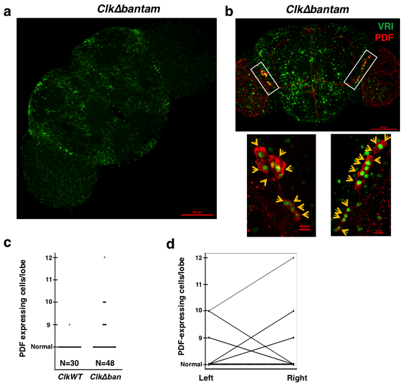
Figure 6. Deletion of the bantam binding sites on the Clk 3’UTR lead to stochastic development of the pdf-expressing cells.
a. Immunofluorescence (IF) analysis of a representative ClkΔban (3-7) Drosophila brain using an anti-VRI antibody. Flies were collected and dissected at ZT15. b. Representative example of a ClkΔban fly brain in which 21 LNvs cells were observed, 9 in one hemisphere and 12 in the other (top panel). Lower panel represents magnification of rectangle area. Of the 12 PDF positive cell bodies in the right hemisphere: 9 cells are VRI positive, one cell display low intensity of VRI immunostaining and 2 cells are VRI negative. In the left hemisphere of the brain, 9 PDF positive cells are observed: 8 cells are VRI positive and one cell displays low intensity of VRI immunostaining. Flies were collected and dissected at ZT15. Arrows indicate the position of each PDF positive cell. c. Number of PDF-positive cell bodies in ClkWT and ClkΔban brains. Data is taken from the ClkΔban (3-7) and ClkWT (1-1) fly strains. d. Comparison of left and right hemispheres in ClkWT and ClkΔban flies.

