Figure 2.

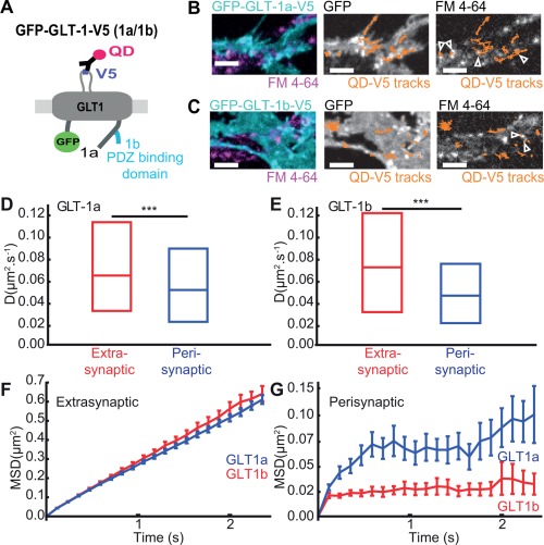
GLT‐1 is stable and confined inside synaptic areas under basal conditions. Astrocytes in hippocampal neuron‐astrocyte mixed culture transfected with GFP‐GLT‐1a‐V5 or GFP‐GLT‐1b‐V5 at DIV10 and imaged at DIV13 after FM4‐64 staining. A: Schematic representation of GFP‐GLT‐1(a/b)‐V5 labelled by an anti‐V5 antibody/QD complex. B,C: Representative time lapse imaging illustrates GFP‐GLT‐1a‐V5 (B) and GFP‐GLT‐1b‐V5 (C) in a region of an astrocyte. GFP‐GLT‐1‐V5 overlaid with FM4‐64 stained synapses (left panels), or by QD‐tagged GFP‐GLT‐1‐V5 trajectories shown in orange (middle panels), and FM4‐64 stained synapses overlaid by QD‐tagged GFP‐GLT‐1‐V5 trajectories shown in orange (right panels), Scale bars 5 μm. D: Instantaneous diffusion coefficients of extrasynaptic GLT‐1a (red, median = 0.065 μm2/s; n = 599 trajectories) and perisynaptic GLT‐1a (blue, median = 0.052 μm2/s; n = 112 trajectories), median D is significantly decreased in perisynaptic areas (P= 6x10 − 3, Mann‐Whitney test). E: Instantaneous diffusion coefficients of extrasynaptic GLT‐1b (red, median = 0.071 μm2/s; n = 255 trajectories) and perisynaptic GLT‐1b (blue, median = 0.047 μm2/s; n = 62 trajectories). Median D is significantly decreased in perisynaptic areas (P = 8 × 10⁻⁵, Mann‐Whitney test). MSDt plot of extrasynaptic (F) and perisynaptic (G) GLT‐1a and GLT‐1b.
