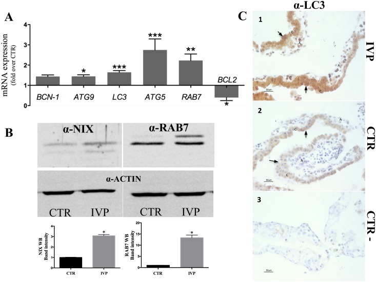
Fig 4. Molecular markers of autophagy in early IVP placentae.
(A) Highly upregulated expression (*p<0.05; **p<0.005; ***p<0.0001) of genes regulating different steps of autophagy in IVP placentae. Note also low expression levels of the antiapoptotic gene, BCL2, which decreases autophagy through BCN-1 interaction. (B) Mitochondria extract from IVP placenta showed an elevated expression of the receptor protein for mitophagy recepror protein NIX. (B) Immunoblotting for canonical marker of autophagy, RAB7 (regulating autophagosome maturation) demonstrated elevated protein expression. (C). Immunostaining of LC3 (marker of autophagy induction) showed an intense and widespread LC3 expression in cells from IVP placentae (1), whereas in CTR placentae LC3 expression seems to be confined to the chorion (2). We used immunohistochemical assay because commercially available antibody against LC3 does not work in sheep under immunoblotting condition. In the figures black arrow shows positive staining of LC3, C denotes the chorion and A indicates the allantois. Control staining without primary antibody (3).

