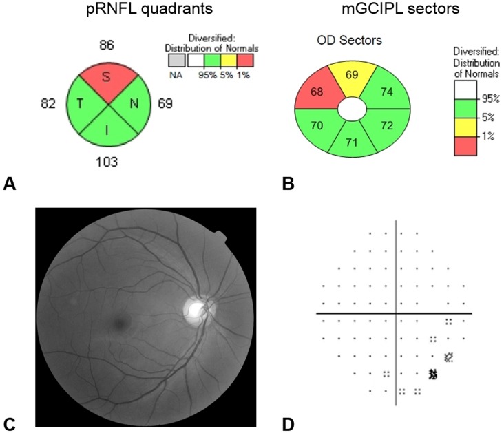
Fig 1. Representative case of anti-SSB positive patient (right eye of 51 year-old female) who showed abnormally reduced thickness of the peripapillary retinal nerve fiber layer (pRNFL) and macular ganglion cell–inner plexiform layer (mGCIPL).
Color coding is as follows: green = normal range, yellow = below the 5th percentile of normal distribution and red = below the 1st percentile of normal distribution. (A) pRNFL thickness below the 5th percentile in average thickness and in at least one segment of the quadrant was considered as an indicator of abnormally reduced pRNFL thickness. (B) mGCIPL thickness below the 5th percentile in average and minimum thickness and in at least one of six sectors was considered as an indicator of abnormally reduced mGCIPL thickness. The patient showed nonspecific findings in RNFL photo (C) and visual field exam (D).

