
Fig 5. Vascularization and Angiogenesis.
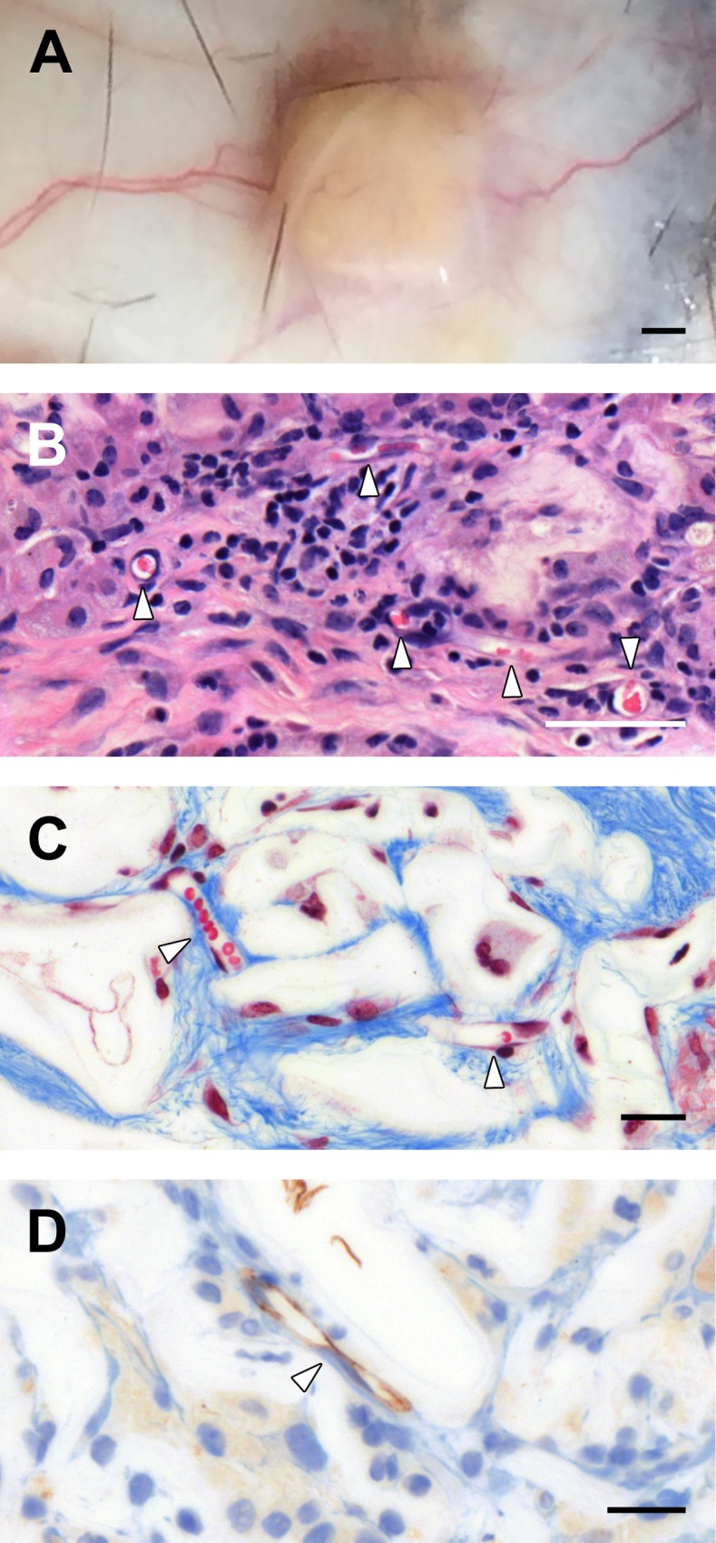
Macroscopic observations of blood vessels directly in the surrounding tissues around the cellulose scaffold (A). Confirmation of angiogenesis within the cellulose scaffold by the observation of multiple blood vessel cross sections in H&E staining (B) and Masson’s Trichrome staining (C) micrographs. The angiogenesis process was also confirmed with anti-CD31 staining to identify endothelial cells within the cellulose scaffold (D). Scale bars: A = 1mm, B = 50μm and C-D = 20μm. White arrows = blood vessels.