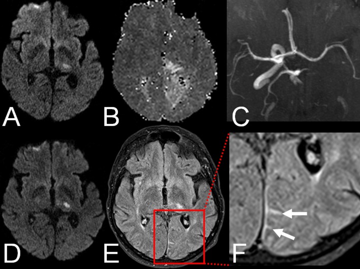
Fig 1.
Example of HARM in a 67-year old patient with left posterior cerebral artery (PCA) infarction: A. Acute ischemic lesion in the left thalamus on DWI. B. Hypoperfusion in the left PCA territory on PWI. C. Proximal occlusion of the left PCA on TOF-MRA. D. Acute ischemic lesion in the left thalamus on follow-up DWI. E. HARM in the left PCA territory on FLAIR images. E. Magnification (1.5x) of HARM on FLAIR images (arrows).

