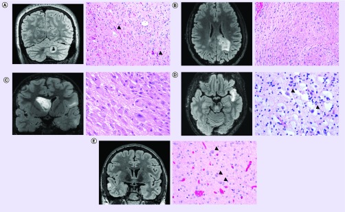
Figure 1. . Representative imaging and histology from pediatric low-grade gliomas.
(A) Pilocytic astrocytoma, WHO grade I, with KIAA1549–BRAF gene fusion. Coronal T2-weighted fluid-attenuated inversion recovery (FLAIR) MRI (left) demonstrating a solid and cystic lesion centered in the cerebellar hemisphere. Hematoxylin and eosin (H&E) stained section (right) demonstrating a compact area of the neoplasm composed of piloid astrocytes containing numerous Rosenthal fibers (examples of which are highlighted by black arrowheads). Molecular analysis (not shown) demonstrated duplication of the 3′ portion of the BRAF gene along with fusion to the KIAA1549 locus. (B) Diffuse astrocytoma, WHO grade II, with MYB gene rearrangement. Axial T2-weighted FLAIR MRI (left) demonstrating an infiltrative hyperintense lesion within the deep white matter of the parietal lobe and infiltrating into the splenium of the corpus callosum. H&E stained section (right) demonstrating infiltrating neoplastic astrocytes with elongate nuclei and coarse chromatin in a background of densely fibrillar glial processes. FISH (not shown) demonstrated rearrangement of the MYB gene in this tumor. (C) Subependymal giant cell astrocytoma, WHO grade I, in a tuberous sclerosis patient. Coronal T2-weighted FLAIR MRI (left) demonstrating an intraventricular mass lesion arising from the caudate nucleus. Also seen are multiple regions of FLAIR-hyperintensity within cerebral cortex and subcortical white matter consistent with cortical tubers. H&E stained section (right) demonstrating a solid neoplasm composed of large epithelioid astrocytes with abundant eosinophilic cytoplasm within a densely fibrillar background. (D) Dysembryoplastic neuroepithelial tumor, WHO grade I. Axial T2-weighted FLAIR MRI (left) demonstrating a well circumscribed, cortically based mass lesion in the temporal lobe with internal nodularity. H&E stained section (right) demonstrating a neoplasm of round oligodendrocyte-like cells arranged in linear columns along capillaries and neuronal processes within a mucin-rich stroma containing ‘floating’ neurons (examples of which are highlighted by black arrowheads). (E) Ganglioglioma, WHO grade I, with BRAF V600E mutation. Coronal T2-weighted FLAIR MRI (left) demonstrating a well circumscribed, cortically based solid and cystic mass lesion in the temporal lobe. H&E stained section (right) demonstrating a biphasic tumor composed of neoplastic glial cells admixed with large dysmorphic ganglion cells (highlighted by black arrowheads). Molecular testing (not shown) demonstrated the presence of BRAF V600E mutation in this tumor.

