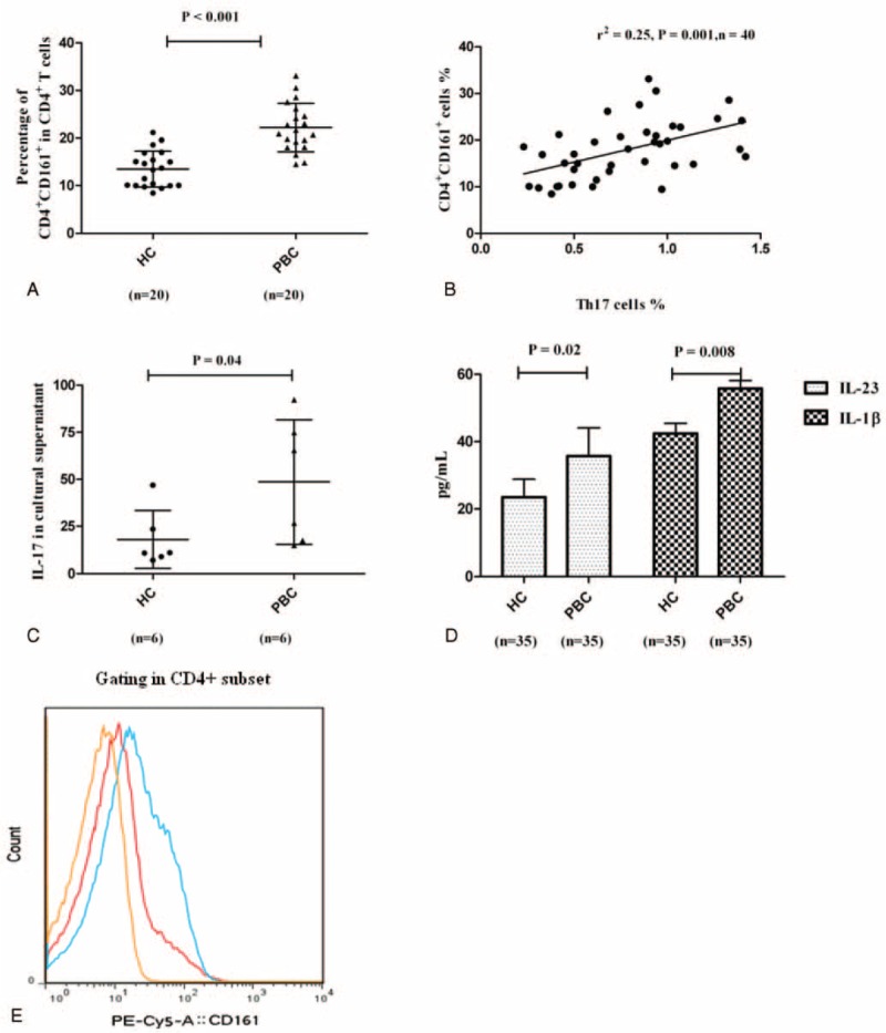
FIGURE 4.

The source for elevated Th17 cells in PBC patients. (A) Frequency of CD161+CD4+ cells in the CD4+ T compartment in PBC patients (n = 20) and HCs (n = 20), respectively. (B) Relationship between Th17 cells and CD161+CD4+ cells (n = 40). (C) IL-17 expression of CD161+CD4+ cells after stimulation in vitro in PBC patients (n = 6) and HCs (n = 6), respectively. (D) Levels of serum IL-23 and IL-1β in PBC patients (n = 35) and HCs (n = 35), respectively. (E) Representative flow cytometry results for CD161+CD4+ cells in the CD4+ T compartment from PBC patients and HCs. HCs = healthy controls, IL = interleukin, PBC = primary biliary cirrhosis, Th = T helper.
