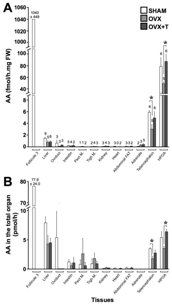
Fig. 9.
Aromatase activity assayed in different peripheral and brain tissues of intact females (SHAM - white), ovariectomized females (OVX – light grey), ovariectomized females treated with testosterone (OVX+T – dark grey). AA was expressed by mg of fresh weight in the three experimental groups (A) or as the total enzymatic activity of the organ in the three experimental groups (B). Numbers above histogram bars in (A) represent the number of samples in which AA was detectable on a total of 6 SHAM, 5 OVX and 6 OVX+T (only 5 for the oviduct because one sample was lost). * p < 0.05. p values are adapted with the Bonferroni correction when appropriate.

