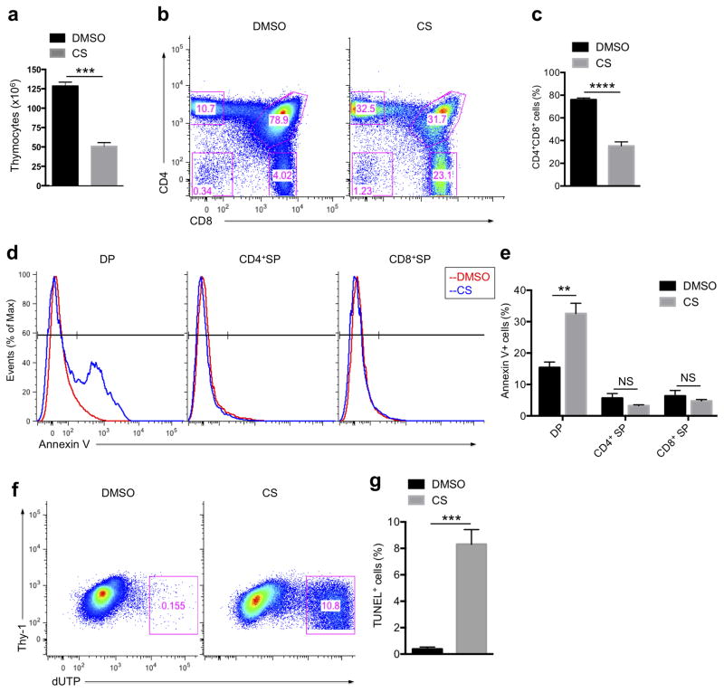
Figure 5. Increasing cholesterol sulfate abundance induces apoptosis in double positive (DP) thymocytes.
(a) Cell number counting of total thymocytes after intrathymic injection of DMSO control or CS. The numbers were estimated using a hemocytometer. (b) Flow cytometry analysis of thymocyte populations. Mice were treated as in a. (c) Statistical analysis for the percentage of DP thymocytes. (d) Flow cytometry analysis of surface exposure of phosphatidylserine. Mice were treated as in a, then thymocytes were subjected to FITC-Annexin V staining. DP (left), CD4+ SP (middle) and CD8+ SP (right) populations were gated and Annexin V staining intensities were analyzed. (e) Statistical analysis for the percentage of Annexin V positive populations. (f) Flow cytometry analysis of TUNEL positive thymocyte population. Mice were treated as in a, then thymocytes were subjected to TUNEL staining and analyzed by flow cytometry. (g) Statistical analysis for the percentage of TUNEL positive populations. **P < 0.01; ***P < 0.001; ****P < 0.0001; NS, not significant, unpaired t-test mean and s.e.m. (a,c,e,g). Data are from one experiment representative of three independent experiments with similar results (b,d,f) or pooled three independent experiments (a,c,g; n = 4, e; n = 5).

