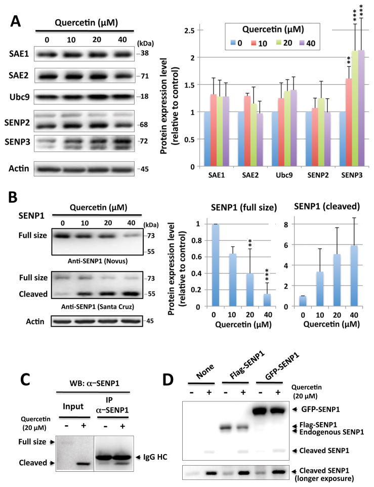
Figure 2. Effects of quercetin on the expression of SUMOylation pathway related proteins.
(A) Representative immunoblots (left) and quantitative analyses (right) of the expression of SUMOylation pathway related proteins: levels of E1 (SAE1, SAE2), E2 (Ubc9) and the isopeptidases (SENP2, SENP3) in SHSY5Y cells that were incubated with quercetin at 0, 10, 20 and 40 μM for 16hr. The density of each band was normalized with the corresponding actin level and expressed as the ratio to control (no quercetin: DMSO alone). (B) Representative immunoblots (left) and quantitative analyses (right) of SENP1 level (full-size and cleaved form). Two SENP1 antibodies, one from Novus Biologicals (NBP1-89553), which was raised against AA 85–205, and one from Santa Cruz Biotechnology (sc-271360) which was raised against AA 361–425 were used. The density of each band was normalized with corresponding actin level and expressed as the ratio to control (no quercetin: DMSO alone). Data represent the mean ± standard deviation of at least three independent experiments. ***p<0.001, **p<0.01, *p<0.05 compared to the DMSO alone control via a one way ANOVA, followed by Dunnett’s post-hoc test. (C) Cell lysates from SHSY5Y cells, which were treated with or without quercetin (20 μM) overnight, were immunoprecipitated with an anti-SENP1 antibody from Santa Cruz. (D) SHSY5Y cells were transfected with either flag-SENP1 or GFP-SENP1, treated with or without quercetin (20 μM) overnight, and the various SENP1 expression levels were examined by the Western blot with anti-SENP1 antibody from Santa Cruz.

