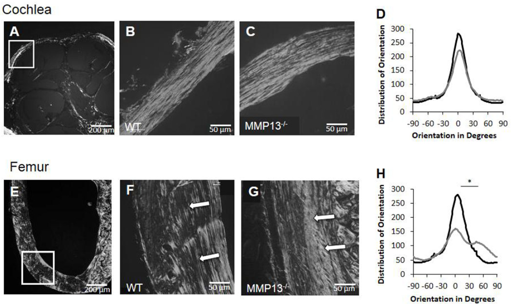
Figure 4. MMP13 is necessary for collagen organization in long bones, not in cochlear bone.
Polarized light birefringence after picro-sirius staining of p60 male mice cochleae and femora reveal collagen fibril organization. Both WT and MMP13−/− cochleae reveal well-organized collagen fibrils (A–C, white box indicates region of interest for panels B–C). Plot of the distribution of orientation of collagen fibrils in cochlear bone (D) reveal no differences between MMP13−/− (36.6±10.4°) and WT mice (33.5±7.2°) by histogram analysis of width at half maximum, p>0.05, via OrientationJ plug-in for ImageJ. In femora (E–G, white box indicates region of interest for panels F–G), collagen organization is not well organized throughout MMP13−/− mice (G) compared to WT (F). For MMP13−/− femora, the distribution of collagen orientation shows a wider, less well-defined peak (H), suggesting poorer organization by width at half-maximum analysis, (71.8±30.6 vs. 34.3±4.8°, MMP13−/− and WT, respectively; p=0.0002). Data from n≥3 wild type mice and n≥3 MMP13−/− mice were obtained.

