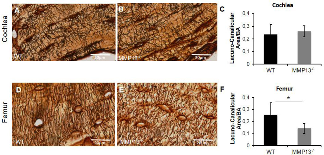
Figure 5. Cochlear canalicular network is maintained without MMP13, however is required in femur.
Lacuno-canalicular networks in cochleae (A–B) and femora (D–E) stained with silver nitrate (in black) in male p60 mice. Silver nitrate highlights the lacunae and the radiating canalicular network in cochlea, with no appreciable difference between WT (A) and MMP13−/− mice (B). Quantification of the lacuno-canalicular area, using ImageJ, reveals no significant difference between the two groups (0.260±0.04µm2 vs. 0.235±0.08µm2, MMP13−/− and WT, respectively, p>0.05) (C). Silver nitrate staining of axial femur section reveals a robust, radiating canalicular network from lacunae in WT femora (D). MMP13−/− mice reveal a less extensive, blunted canalicular network (E). Quantified via ImageJ, MMP13−/− mice have a significantly smaller lacuno-canalicular area (0.143±0.04 µm2) normalized to bone area, compared to WT mice (0.256±0.10 µm2; p=0.0002) (F). Data was collected from n≥3 cochleae from each group, and from n≥3 MMP13−/− and n=2 wild type mice femora.

