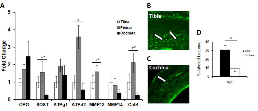
Figure 6. Critical PLR factors are expressed to a lesser degree in the cochlea compared to long bones.
(A) qPCR expression levels from p60 WT male mice of osteoprotegerin (OPG), sclerostin (SOST), ATPase H+ transporting lysosomal V0 subunit D2 (ATP6V0D2), ATPase H+ transporting lysosomal V1 subunit G1 (ATP6V1G1), matrix metalloproteinase-13 (MMP13), matrix metalloproteinase-14 (MMP14), and cathepsin-K (CatK) mRNA in cochlea and femur normalized to GAPDH, standardized to levels in the tibia. The relative expression levels of MMP13 and other important PLR genes (CatK, ATPd2, MMP14) in the cochlea are lower than in other bones, suggesting suppressed PLR activity in the cochlea relative to long bones (“+” indicates normalized to tibia, “*” indicates normalized to femur). This is reinforced by quantifying the percentage of calcein labeled lacunae in WT tibia and WT cochlea (D), where tibia revealed 31% labeled lacunae versus 9% in the cochlea (p-value=0.00002). Representative images under confocal microscopy of calcein-stained tibia (B) showing a relatively high number of lacunae with calcein label (Tibia, white arrows) and the relative paucity in cochlea (C) (Cochlea, white arrow). Data was collected from bones from n=3 mice per group.

