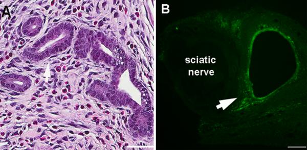
Figure 2.
Grafts had histological features of uterine tissue. Examples of cross sections of the sciatic nerve and adjacent uterine graft on POD21. A: H&E staining showed characteristic gland formations with columnar cells such as are seen in normal uterus. Scale bar = 50μm. B: Staining for cytokeratin-18, a marker for glandular epithelial cells. Note that the marker is not seen inside the sciatic nerve. Arrows indicate labeled tissue. Scale bar = 100μm.

