Figure 5.
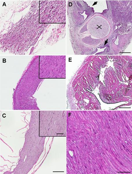
H&E staining of nerve and uterine graft showed signs of inflammation. A. Longitudinal section of nerve adjacent to uterine implant on POD21. Note inflammatory cell infiltration, swelling leading to spaces between nerve fibers, and destruction of nerve fibers. B. Longitudinal section of nerve adjacent to uterine graft on POD49. Fewer inflammatory cells and amelioration of swelling between nerve fibers were observed. C. Inflammatory signs disappeared when the implant was removed on POD21 (image taken 14 days after removal). Longitudinal section of nerve adjacent to uterine implant. D. Cross-section of sciatic nerve adjacent to uterine graft showing sciatic nerve (x) and endometrium (arrows) around the sciatic nerve on POD21. Note extensive inflammatory cell infiltration of the endometrium. E. Cross section of uterine implant tissue on POD49. Fewer inflammatory cells were observed. F. Longitudinal section of nerve adjacent to fat tissue implant on POD21 does not show inflammation or nerve damage. Figure A–E under 4× objective, upper right areas and figure F under 20× objective. Scale bar A–E = 400μm, upper right areas and F = 70μm.

