
Fig. 2.
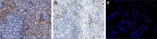
Immunohistochemical (IHC), in situ mRNA and fluorescence in situ hybridization (FISH) analyses of PD-L1 protein expression and CD274 gene in patients with triple-negative breast cancer (TNBC). a Expression of PD-L1 protein in both immune and tumor cells (score as 3+) (×200); b In situ mRNA hybridization of PD-L1 from the same patient is indicated by brown staining (×200); c Representative image obtained from the same patient shows a copy number gain in CD274 gene, with CD274 (green) and CEN9 (red) on chromosome 9p24.1 (×1000)