
Figure 4.
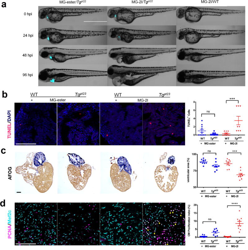
FAP-TAPs induced photo-ablation of cardiac function. (a) Phenotype development from 0 hpi to 96 hpi of larval zebrafish (Merge of DIC and mCer3 fluorescence (cyan), n = 20 for each group): MG-ester/Tgpt22, MG-2I/Tgpt22 and MG-2I/WT. In MG-2I/Tgpt22 group, the larvae developed a range of visible defects: large cardiac edema, small eyes, and collapsed, nonfunctional heart chambers. In both control groups, development proceeded normally. Scale Bar = 1000 μm and applied to all images; (b) FAP-TAPs photo-induced cardiac damage in adult zebrafish. Hearts were extracted at 3 dpi and TUNEL was performed to assess cell death; (c) Acid Fuchsin Orange G (AFOG) staining of hearts at 5 dpi showing the damaged cardiac structure in transgenic fish injected with MG-2I; (d) Mef2c (cardiomyocyte) and PCNA (proliferation) staining at 5 dpi shows enhanced cardiomyocyte proliferation (yellow arrow) in transgenic fish injected with MG-2I. Scale bar = 100 μm and applied to all images, n = 9 for all groups, One-way ANOVA, Tukey post hoc tests were performed with multiple comparisons of mean for each group. P-values were considered significant when < 0.05, shown as mean ± S.E.M.