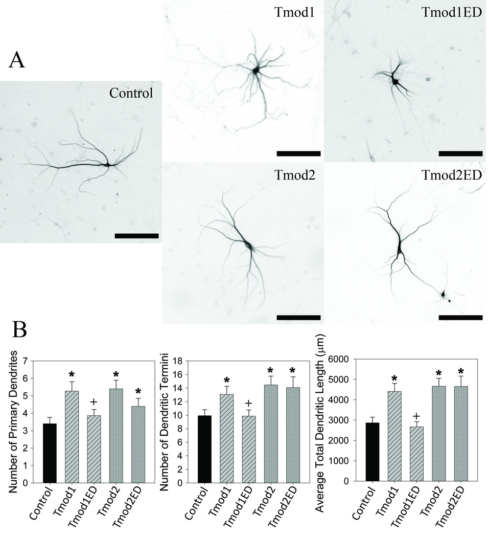
Figure 4. Tpm binding is necessary for Tmod1’s but not for Tmod2’s impact on dendritic morphology.
Primary hippocampal neurons were transfected with pCAGGs plasmid encoding ClFP-tagged Tmod, wild type or with disrupted Tpm-binding sites. A. Representative images of neurons analyzed in this experiment. Shown images are RFP signals from RFP-MAP2b used for morphology analysis (scale bars = 150 µm). B. Imaged neurons were analyzed for number of primary dendrites (left), dendritic termini (center) and total dendritic length (right). 15–22 neurons were analyzed for each condition. Error bars indicate SEM. Asterisks and plus symbols indicate statistically significant difference from controls and wild type overexpression, respectively (ANOVA, P < 0.05).

