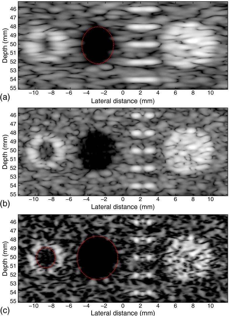
Fig. 7.
Field II simulated images of a phantom. Conventional B-scan using (a) Gaussian and (b) rectangular apodization with focus at about 50 mm. (c) Enhanced result. The lateral diameter of the cyst is increased from 3.7 to 4.7 mm, and the blood vessel wall (left) is seen much more clearly than that in the original image. The diameter of the inner blood vessel wall, which is designed to be 3 mm, is opened from barely visible to about 2.1 mm. The scatterer pairs are separated after inverse filtering.

