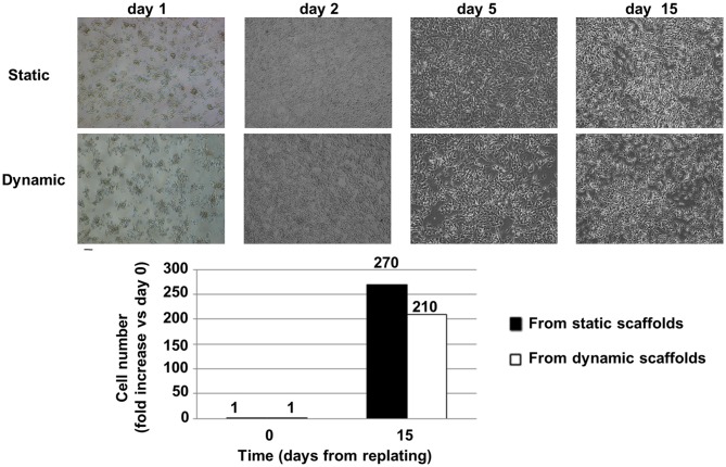
Figure 6.
Cell recovery from the bioreactor and replating. Top: Brightfield images of cell morphology after replating: static vs. dynamic (48 h in the 4D bioreactor). SH-SY5Y cells were carefully detached from 3D scaffolds and plated in standard 2D conditions in a 12-well microplate (day 0 was assumed as the day of replating). On day 2 cells were moved to a T25 cm2 flask, on day 5 they were further expanded in T25 cm2 flasks, while on day 15 their serum-free conditioned culture medium was collected from T75 cm2 flasks for exosome isolation. Scale bar = 100 μm. Bottom: Fold increase (mean values) in cell number with respect to the day of replating. Cells were counted after detachment with trypsin.

