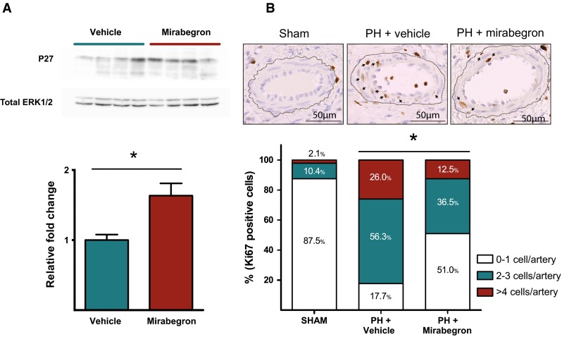
Fig. 2.

Protein expression related with pulmonary cellular proliferation. a Western blot for the P27 protein in the lung parenchyma from pigs with chronic PH receiving vehicle (N = 4) or mirabegron (N = 4) for 14 days. The densitometric analysis of P27 normalized to total ERK 1/2 is shown below. b Representative immunohistochemical pictures for Ki67 staining in pulmonary arteries within the lung parenchyma from sham-operated controls, pigs with chronic PH treated with vehicle and pigs with chronic PH treated with mirabegron. Brown staining indicates Ki67-positive cells. Arrows indicate Ki67-positive cells within the arterial wall. The frequency bar chart shown below represents the percentage of pulmonary arteries with Ki67 positive cells categorized in three groups (0–1 positive cell/artery, 2–3 positive cells/artery and >4 positive cells per artery) by group. *Statistically significant differences
