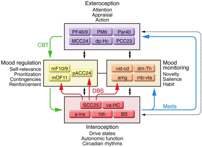
Figure 2.

A schematic representation of the network of connections between cortical, limbic, and paralimbic regions proposed to underlie major depressive disorder (MDD), and the four main functional compartments of the model, each of which represents a functional domain impaired in MDD and comprises a subset of brain regions that demonstrate robust anatomical connections to one another. Black arrows identify anatomical connections between compartments. Solid colored arrows identify connections that are proposed to mediate efficacy of a specific treatment: green indicates cognitive behavioral therapy; blue indicates pharmacotherapy; red indicates deep brain stimulation (DBS) at subcallosal cingulate. Additional abbreviations used in the figure: a-ins, anterior insula; amg, amygdala; dm-Th, dorsomedial thalamus; dp-Hc, dorsal-posterior hippocampus; mb-vta, midbrain-ventral tegmental area; mF10/9, medial frontal cortex BA10 and BA9; mOF11, medial orbital frontal cortex BA11; Par40, parietal cortex BA40; PF46/9, PFC BA46 and BA9; PM6, premotor cortex BA6; va-HC, ventral-anterior hippocampus; vst-cd, ventral striatum-caudate. Republished with permission of American Society for Clinical Investigation, from Mayberg (2009); permission conveyed through Copyright Clearance Center, Inc.
